



Contents
Scroll to:
https://doi.org/10.17749/2313-7347/ob.gyn.rep.2026.677
Scroll to:
Introduction. The rising global incidence of cesarean deliveries has led to elevated abnormal placentation, particularly placenta accreta spectrum (PAS) disorders. These conditions represent serious obstetric complications with high risks for both maternal and fetal morbidity and mortality.
Аim: to analyze a clinical case of placenta increta with deep invasion, highlighting the diagnostic value of advanced ultrasonographic techniques.
Case presentation. A 29-year-old pregnant woman with a history of three cesarean sections (2014, 2017, and 2025) and prominent somatic comorbidities (chronic pyelonephritis and urolithiasis) underwent routine antenatal ultrasound screenings at 8, 12, 20, 23, and 28+6 weeks of gestation. Standard transvaginal and transabdominal sonographic techniques were used. After sonographic suspicion of placenta increta, magnetic resonance imaging (MRI) was performed for verification. Final diagnosis was confirmed intraoperatively and through histopathological analysis. Ultrasound revealed signs consistent with PAS type 2 (placenta increta): marked thinning of the myometrium, dilated lacunae, retroplacental hypervascularization, and placental bulging. At 28+6 weeks, transvaginal sonography detected further invasion into the cervical canal (PAS 3a). Color Doppler imaging revealed vascular "rail signs," indicating deep invasion. MRI confirmed complete placenta previa with abnormal tissue growth into the uterine scar and myometrial layers. This case demonstrates the effectiveness of modern ultrasonography and Doppler imaging in the early identification and classification of PAS disorders. Although previous publications emphasized limitations in determining invasion depth via ultrasound, this report highlights its diagnostic reliability, especially when complemented by MRI. Early diagnosis is essential for surgical planning and improving perinatal outcomes.
Conclusion. Ultrasound primarily combined with color Doppler imaging, provides a reliable and non-invasive means for diagnosing PAS disorders. In this case, the identification of specific Doppler features – particularly the "rail sign" representing vascular bridging between the myometrium and serosa – proved critical for diagnosing placenta increta. These findings highlight a potential for advanced Doppler markers in improving antenatal detection and surgical planning. Integrating such diagnostic techniques into routine care can markedly improve maternal and fetal outcomes in high-risk pregnancies.
Eliava E., Verulava T. Role of modern ultrasonography in antenatal detection of placenta increta: a case report. Obstetrics, Gynecology and Reproduction. 2026;20(1):220-228. https://doi.org/10.17749/2313-7347/ob.gyn.rep.2026.677
Placenta accreta is a severe pregnancy complication that occurs when placental tissue and blood vessels abnormally invade the uterine wall. The history of placenta accreta dates back more than a century. The earliest case documented in 1890 is reported from Tübingen, Germany, in 1890. Later, in 1937, C. Irving and A.T. Hertig published the first comprehensive case series, providing a systematic description and classification of abnormal placental adherence [1]. While earlier single case reports existed, their work laid the foundation for clinical recognition and standardization of the diagnosis.
In recent years, the terminology and classification of this pathology was revised. The term Placenta Accreta Spectrum (PAS) is now globally accepted and encompasses a range of abnormal placental adherence and invasion, including placenta accreta, increta, and percreta [2]. PAS disorders have become more prevalent alongside the global rise in cesarean deliveries, posing serious risks to both maternal and fetal health.
PAS disorders are multifactorial in origin and are strongly associated with defective decidualization and excessive trophoblastic invasion. Normally, the decidua basalis provides a protective barrier that prevents chorionic villi from invading the myometrium. When this barrier lacks or underdeveloped, commonly due to previous uterine surgeries such as cesarean section, myomectomy, or curettage, the placenta may penetrate the myometrium pathologically [3].
Established risk factors for PAS include:
These factors often overlap, increasing the overall risk. In the current case, the patient's history of multiple cesarean deliveries, placenta previa, and chronic urological conditions referred her to a high-risk category. Identifying such risk factors is essential for early diagnosis, appropriate imaging, and planning delivery in specialized centers.
The traditional PAS classification used widely for many years is based on assessing the depth of chorionic villi invasion [4]:
The currently accepted PAS classification was proposed by the International Federation of Gynecology and Obstetrics (FIGO), which includes [5]:
The precise PAS etiopathogenesis remains under investigation. One hypothesis links PAS to defective decidual formation (e.g., lacked decidua basalis or underdeveloped fibrinoid layer), abnormal vascular remodeling during pregnancy, and excessive trophoblastic invasion, particularly following prior uterine surgery.
Placental adherence may be:
Over the past four decades, cesarean section rates have risen by over 20 %, while PAS cases have increased tenfold [6]. The prevalence of PAS varies between 1 in 500 to 1 in 10,000 deliveries, with complete placental invasion being particularly rare – around 1 in 24,506 births [7][8]. Among PAS cases: accreta accounts for 60–75 %, increta for 15–20 %, and percreta for 5–10 %.
Despite ultrasound being the primary tool for PAS detection, distinguishing between placenta accreta, increta, and percreta remains challenging using grayscale imaging alone. Depth of invasion is difficult to assess due to overlapping sonographic features and variation in interpretation. Classic markers such as placental lacunae, myometrial thinning, and loss of the retroplacental clear zone are helpful but not specific, especially in early gestation.
Color Doppler imaging and emerging signs such as the “rail sign” (vascular bridging between placenta and adjacent tissues) improve diagnostic precision. However, these findings are not yet standardized and require operator experience and high-resolution equipment. Likewise, advanced tools like 3D power Doppler and shear-wave elastography are being explored but are not yet widely adopted into routine clinical protocols. Unlike most previously reported cases, current report highlights the diagnostic contribution of the "rail sign" on color Doppler as an early marker of placenta increta, verified later by MRI. To our knowledge, such a detailed correlation between Doppler vascular markers and intraoperative findings has been rarely documented.
The present case contributes to this field by demonstrating the practical value of vascular Doppler markers for diagnosing placenta increta and early signs of PAS 3a.
Historically, PAS was only diagnosed postnatally upon complicated placental separation or postpartum hemorrhage, as seen in early reports by Forster, Irving, and Hertig. Today, ultrasound is the primary modality for antenatal detection [9–12].
The meta-analysis by D’Antonio et al. assessing 3,070 high-risk pregnancies, demonstrated that ultrasound sensitivity and specificity comprised 90.72 % and 96.94 %, respectively, for PAS diagnosis [13].
Sonographic indicators of PAS include [14][15]:
Key Doppler criteria include [10][11]:
Early diagnosis allows for planned delivery in tertiary centers, reduces emergency hysterectomy rates, and improves maternal and neonatal outcomes. While cases of antenatal PAS diagnosis are well-documented globally, such reports are limited in Georgian literature. This article presents a clinical case that illustrates the role of modern ultrasound, particularly Doppler techniques, in the diagnosis and management of placenta increta.
Patient D., 29 years old, has a history of three cesarean deliveries (in 2014, 2017, and 2025). The somatic history is significant for chronic pyelonephritis and urolithiasis.
The patient underwent routine screening examinations at 8, 12, 20, 23, and 28⁺⁶ weeks of gestation. Standard transvaginal and transabdominal ultrasounds were performed. After diagnosing placenta increta, magnetic resonance imaging (MRI) was additionally conducted. The diagnosis was confirmed intraoperatively and by histopathological examination.
At gestational age of 8 weeks, first-degree invasion of the gestational sac into the scar area was observed.
At 12–13 weeks of gestation, the placenta was localized on the anterior uterine wall, in the lower segment, covering the medial aspect of the postoperative scar and extending to the internal os of the endocervical canal – indicating placenta previa (Fig. 1).
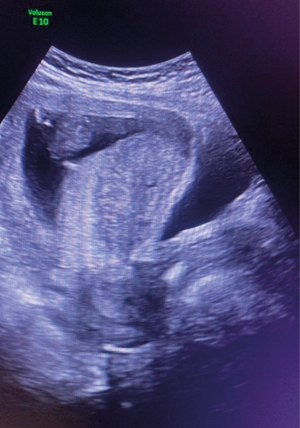
Figure 1. Patient D. at 12–13 weeks of pregnancy.
Рисунок 1. Пациентка Д. на 12–13‑й неделе беременности.
During the 12-week screening, both placenta previa and partial invasion into the residual myometrium were identified. The patient was classified as high-risk for placental implantation anomalies and was informed about potential complications (Fig. 2).

Figure 2. Patient D. at 15–16 weeks of pregnancy.
Рисунок 2. Пациентка Д. на 15–16‑й неделе беременности.
At 19 weeks of gestation, the ultrasound revealed placenta previa localized over the anterior uterine wall at the site of the previous cesarean scar. The myometrial thickness over the scar was 3.2 mm, indicating its significant thinning. Multiple irregular placental lacunae, ranging in size from 4 to 10 mm, were observed with chaotic flow patterns (Fig. 3). Color Doppler examination showed hypervascularity in the retroplacental and bladder interface zone. Peak systolic velocities exceeded 20 cm/s, with low-resistance arterial and venous flow consistent with abnormally invasive vasculature (Fig. 4).
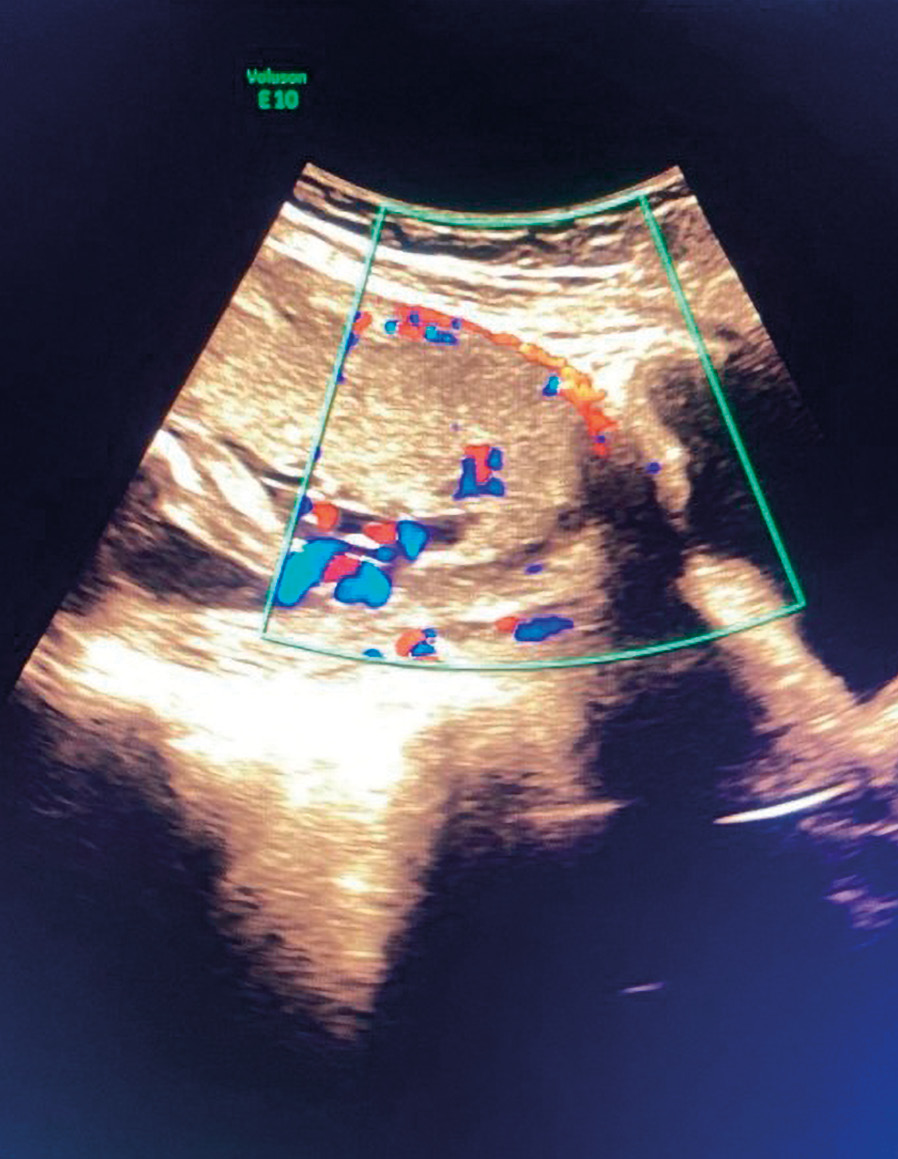
Figure 3. Patient D., 20 weeks of pregnancy.
Рисунок 3. Пациентка Д., 20 недель беременности.

Figure 4. Patient D., 24 weeks of pregnancy.
Рисунок 4. Пациентка Д., 24 недели беременности.
By 28⁺⁶ weeks of gestational age, placental bulging and invasion into the cervical canal were documented, consistent with PAS 3a. The Doppler “rail sign,” characterized by parallel bridging vessels extending from the myometrium through the serosa, was clearly visualized.
At 24 weeks of gestation, placenta previa was noted again. The myometrial structure could not be assessed as the placenta was fully invading it; the serosal layer remained intact, the endocervical canal was closed, with its length was 40 mm. According to the FIGO classification, this corresponded to PAS 2.
At 28⁺⁶ weeks of gestational age, transvaginal scanning revealed invasion of the placenta into the upper third of the cervix and the cervical canal corresponding to PAS 3a (Table 1).
Table 1. Timeline summary of ultrasound and Doppler parameters in placenta accreta spectrum (PAS) diagnosis.
Таблица 1. Хронология ультразвуковых и доплеровских параметров при диагностике placenta accreta spectrum (PAS).
|
Gestational age Гестационный возраст |
Findings Результаты |
Myometrial thickness Толщина миометрия |
Lacunae Лакуны |
Doppler velocity Доплеровская скорость |
PAS classification Классификация PAS |
|
8 weeks 8 недель |
Suspected scar invasion Подозрение на инвазию в рубец |
Not measurable Неизмеримо |
Not present Отсутствуют |
Not assessed Не оценивалась |
– |
|
12 weeks 12 недель |
Partial intra-scar invasion Частичная инвазия в рубец |
~4 mm ~4 мм |
Small lacunae Небольшие лакуны |
Mild flow Слабое течение |
Suspected PAS 1–2 Подозрение на PAS 1–2 |
|
19 weeks 19 недель |
Placenta previa, hypervascularity Предлежание плаценты, гиперваскуляризация |
3.2 mm 3,2 мм |
Multiple (4–10 mm) Множественные (4–10 мм) |
> 20 cm/s > 20 cм/с |
PAS 2 PAS 2 |
|
28⁺⁶ weeks 28⁺⁶ недель |
Cervical invasion, “rail sign” Инвазия в шейку матки, «рельсовый симптом» |
Thinned, indistinct Истонченный, нечеткий |
Enlarged lacunae Увеличенные лакуны |
Persistent turbulent flow Постоянное турбулентное течение |
PAS 3a PAS 3а |
Additional Doppler imaging allowed to identify vascular bridges (rail sign) extending from the myometrium through the serosal layer, defined as placenta increta (PAS 2).
Subsequent MRI confirmed complete placenta previa, with abnormal growth into the uterine scar and across all layers of the myometrium.
Doppler evaluation of blood flow proved to be a valuable tool for assessing the degree of abnormal placental invasion.
This clinical case demonstrates the effectiveness of modern ultrasonographic technologies, particularly color Doppler imaging, in diagnosing deep placental invasion. Although Jauniaux et al. previously emphasized the limitations for ultrasound in assessing the depth of placenta increta, this case challenges that viewpoint [16].
The MRI findings corroborated the Doppler-based suspicion of placenta increta with cervical extension (PAS 3a), confirming the depth of myometrial invasion and its relationship to adjacent structures [17]. The diagnostic features, such as myometrial thinning, enlarged lacunae, and abnormal vascular bridging (“rail sign”), are consistent with the FIGO classification for PAS and align with Jauniaux et al.’s standardized ultrasound descriptors. Furthermore, the approach is consistent with the Society for Maternal-Fetal Medicine (SMFM) recommendations, which emphasize the use of grayscale and Doppler imaging for antenatal diagnosis and risk stratification.
While the FIGO classification provides a standardized framework for defining PAS disorders, it has notable limitations. Several specialists have argued that its reliance on histopathological criteria offers little value intraoperatively, when real-time surgical decisions are required. Moreover, studies have highlighted that the FIGO grading does not correlate consistently with maternal morbidity, which limits its prognostic utility. In clinical practice, classifications based on surgical and imaging findings, such as those proposed by Jauniaux et al. (2019) or SMFM, may be more useful, as they emphasize parameters that directly influence management planning, surgical complexity, and outcomes [18]. Incorporating these clinically oriented frameworks alongside FIGO may therefore provide a more comprehensive approach for risk stratification and operative preparation.
We recognize that antenatal ultrasound PAS diagnosis has been well established in the literature. However, the presented case adds more value by demonstrating the utility of the "rail sign" in early gestation and confirming its correlation with MRI and histopathology, thereby reinforcing the clinical applicability of this marker in routine practice.
An additional limitation relates to the pathological assessment per se. Histopathological confirmation is traditionally regarded as the gold standard for PAS diagnosis; however, its validity has been questioned. The degree of placental adherence can vary even within the same specimen, and a diagnosis of placenta increta or percreta may be influenced by the site from which tissue is sampled. This introduces the risk of sampling bias and may lead to under- or over-classification of PAS severity. For this reason, intraoperative findings and the overall clinical context should be considered alongside histological results while establishing a definitive diagnosis.
Despite its high sensitivity and specificity, ultrasound (even when combined with color Doppler or MRI) cannot fully predict the intraoperative degree of surgical difficulty. Numerous studies have shown discrepancies between antenatal imaging findings and actual operative challenges, particularly regarding the extent of vascular invasion, adhesion severity, and risk of hemorrhage. Therefore, while imaging plays a critical role in early detection and management planning, it should not be overestimated as a predictor of surgical complexity. Multidisciplinary readiness and intraoperative adaptability remain essential in managing PAS cases.
Early detection of PAS, especially placenta percreta variants, is vital for maternal and fetal survival, allowing for planned surgical management in high-resource settings. This case illustrates how the integration of Doppler markers into standard prenatal surveillance can improve outcomes and reduce a need for emergency interventions. While limited to a single patient, the findings presented contribute to the growing body of evidence supporting the role for advanced ultrasound in PAS diagnosis. Further studies are needed to validate these vascular markers across diverse clinical settings.
Over the past four decades, the rise in placenta accreta cases has underscored the urgent need for timely and accurate diagnosis of this high-risk pregnancy complication. The growing prevalence of PAS disorders parallels the global increase in cesarean deliveries. Current classification systems, such as the FIGO framework, have facilitated systematic diagnosis and clinical management of PAS variants.
This clinical case reinforces the value of ultrasound, including transvaginal scanning and Doppler imaging, as effective, accessible tools for evaluating the depth of placental invasion. Notably, the use of Doppler-based vascular signs, such as the “rail sign,” provided early indicators of placenta increta with cervical extension (PAS 3a), which were later verified by MRI. This case illustrates how the integration of advanced ultrasound technologies can markedly enhance diagnostic accuracy.
Early PAS identification allows for planned delivery in specialized centers, reducing maternal morbidity and emergency surgical interventions. The presented case underscores how incorporating Doppler markers into routine screening of high-risk patients can influence clinical decision-making, including the timing of delivery, surgical preparation, and maternal outcome optimization. Continuous ultrasonographic monitoring in high-risk pregnancies and incorporation of novel imaging markers into routine protocols may improve perinatal outcomes and guide future PAS management strategies.
While based on a single case, these findings highlight the promise of combining Doppler sonography with anatomical imaging, supporting future research into diagnostic standardization and validation.
1. Irving C., Hertig A.T. A study of placenta accreta. Surg Gynecol Obstet. 1937;64:178–200.
2. Morlando M., Collins S. Placenta accreta spectrum disorders: challenges, risks and management strategies. Int J Womens Health. 2020;12:1033–45. https://doi.org/10.2147/IJWH.S224191.
3. American College of Obstetricians and Gynecologists; Society for Maternal-Fetal Medicine. Obstetric care consensus no. 7: placenta accreta spectrum. Obstet Gynecol. 2018;132(6):e259–e275. https://doi.org/10.1097/AOG.0000000000002983.
4. Jauniaux E., Bhide A. Prenatal ultrasound diagnosis and outcome of placenta previa accreta after cesarean delivery: a systematic review and meta-analysis. Am J Obstet Gynecol. 2017;217(1):27–36. https://doi.org/10.1016/j.ajog.2017.02.050.
5. Collins S.L., Ashcroft A., Braun T. et al. Proposal for standardized ultrasound descriptors of abnormally invasive placenta (AIP). Ultrasound Obstet Gynecol. 2016;47(3):271–5. https://doi.org/10.1002/uog.14952.
6. Sokheim K.N., Esakoff T.F., Little S.E. et al. The effect of cesarean delivery rates on the future incidence of placenta previa, placenta accreta, and maternal mortality. J Matern Fetal Neonatal Med. 2011;24(11):1341–6. https://doi.org/10.3109/14767058.2011.553695.
7. Armstrong C., Harding S., Matthews T., Dickinson J.E. Is placenta accreta catching up with us? Aust N Z J Obstet Gynaecol. 2004;44(3):210–3. https://doi.org/10.1111/j.1479-828X.2004.00208.x.
8. Chalubinski K.M., Pils S., Klein K. et al. Prenatal sonography can predict degree of placental invasion. Ultrasound Obstet Gynecol. 2013;42(5):518–24. https://doi.org/10.1002/uog.12451.
9. Ungiadze J.Y., Nikuradze I.V., Zamtaradze N.D. The role of prenatal diagnosis of abnormally invasive placenta in pregnancy outcome. [Rol' prenatal'noj diagnostiki anomal'no invazivnoj placenty v iskhode beremennosti]. Obstetrics, Gynecology and Reproduction. 2020;14(3):384–94. (In Russ.). https://doi.org/10.17749/2313-7347/ob.gyn.rep.2020.151.
10. Voronina M.D., Dolgiev D.A., Dolgieva R.A. et al. Current state of the placenta accreta spectrum issue: prospects for organ-preserving treatment. [Sovremennoe sostoyanie problemy prirashcheniya placenty: perspektivy organosohranyayushchego lecheniya]. Obstetrics, Gynecology and Reproduction. 2024;19(1):82–96. (In Russ.). https://doi.org/10.17749/2313-7347/ob.gyn.rep.2024.571.
11. Jauniaux E., Collins S.L., Jurkovic D., Burton G.J. Accreta placentation: a systematic review of prenatal ultrasound imaging and grading of villous invasiveness. Am J Obstet Gynecol. 2016;215(6):712–21. https://doi.org/10.1016/j.ajog.2016.07.044.
12. Jauniaux E., Alfirevic Z., Bhide A.G. et al.; Royal College of Obstetricians and Gynaecologists. Placenta praevia and placenta accreta: diagnosis and management. Green-top Guideline № 27a. BJOG. 2019;126(1):e1–e48. https://doi.org/10.1111/1471-0528.15306.
13. D'Antonio F., Iacovella C., Bhide A. Prenatal identification of invasive placentation using ultrasound: systematic review and meta-analysis. Ultrasound Obstet Gynecol. 2013;42(5):509–17. https://doi.org/10.1002/uog.13194.
14. Comstock C.H. Antenatal diagnosis of placenta accreta: a review. Ultrasound Obstet Gynecol. 2005;26(1):89–96. https://doi.org/10.1002/uog.1926.
15. Shih J.C., Palacios Jaraquemada J.M., Su Y.N. et al. Role of threedimensional power Doppler in the antenatal diagnosis of placenta accreta: comparison with gray scale and color Doppler techniques. Ultrasound Obstet Gynecol. 2009;33(2):193–203. https://doi.org/10.1002/uog.6284.
16. Jauniaux E., Ayres-de-Campos D., Langhoff-Ross J. et al.; FIGO Placenta Accreta Diagnosis and Management Expert Consensus Panel. FIGO classification for the clinical diagnosis of placenta accreta spectrum disorders. Int J Gynecol Obstet. 2019;146(1):20–4. https://doi.org/10.1002/ijgo.12761.
17. Lim P.S., Greenberg M., Edelson M.I. et al. Utility of MRI in the diagnosis and management of placenta accreta spectrum disorders: A systematic review. AJR Am J Roentgenol. 2011;197(2):468–75. https://doi.org/10.2214/AJR.11.6858.
18. Jauniaux E., Kingdom J.C., Silver R.M. A comparison of recent guidelines in the diagnosis and management of placenta accreta spectrum disorders. Best Pract Res Clin Obstet Gynaecol. 2021;72:102–16. https://doi.org/10.1016/j.bpobgyn.2020.06.007.
Ekaterine Eliava, MD
Scopus Author ID: 57205196899
66 Lubliana Str., 0159 Tbilisi; 13 Chkondideli Str., Senaki 4100
Tengiz Verulava, MD, Dr Sci Med, Prof.
Scopus Author ID: 56602860500
1 Paata Saakadze Str., 0102 Tbilisi; 124 La Trobe Street Melbourne 3000
What is already known about this subject?
► Placenta accreta spectrum (PAS) disorders are strongly associated with prior cesarean delivery and placenta previa, with incidence rising in parallel with increasing cesarean section rates.
► Ultrasound, especially with color Doppler, is the primary imaging modality for antenatal PAS diagnosis; however, differentiating between placenta accreta, increta, and percreta remains challenging.
► Magnetic resonance imaging (MRI) serves as a complementary tool in complex or inconclusive cases, particularly in posterior placenta or suspected deep invasion into adjacent organs.
What are the new findings?
► Early identification of the “rail sign” on color Doppler provided an initial marker of placenta increta, later confirmed by MRI and histopathology.
► Serial ultrasound monitoring from 8 to 28+6 weeks of gestation documented the progression of placental invasion, offering practical guidance for clinicians managing high-risk pregnancies.
► The integration of Doppler markers with MRI findings reinforced diagnostic accuracy, demonstrating their combined value in planning delivery strategies and improving maternal outcomes.
How might it impact on clinical practice in the foreseeable future?
► Routine incorporation of Doppler markers, such as the “rail sign,” into PAS screening may allow earlier and more reliable detection of abnormal placentation.
► Serial ultrasound monitoring protocols could improve risk stratification, guide delivery planning, and reduce emergency interventions in high-risk pregnancies.
► Combining ultrasound and MRI findings may establish a more standardized diagnostic route, optimizing surgical preparedness and improving maternal-fetal outcomes.
Eliava E., Verulava T. Role of modern ultrasonography in antenatal detection of placenta increta: a case report. Obstetrics, Gynecology and Reproduction. 2026;20(1):220-228. https://doi.org/10.17749/2313-7347/ob.gyn.rep.2026.677


 Email this article (Login required)
Email the author (Login required)
Email this article (Login required)
Email the author (Login required) 





